
Neurological Examination: A Complete Guide for Nursing Students
The human nervous system controls movement, sensation, memory, speech, balance and many essential functions of the body. When a person develops symptoms such as a severe headache, weakness, numbness, confusion or difficulty speaking, a neurological examination can help healthcare professionals assess nervous system function and identify signs that require further investigation.
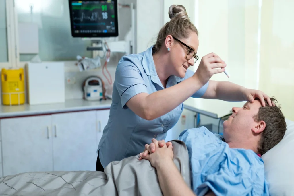
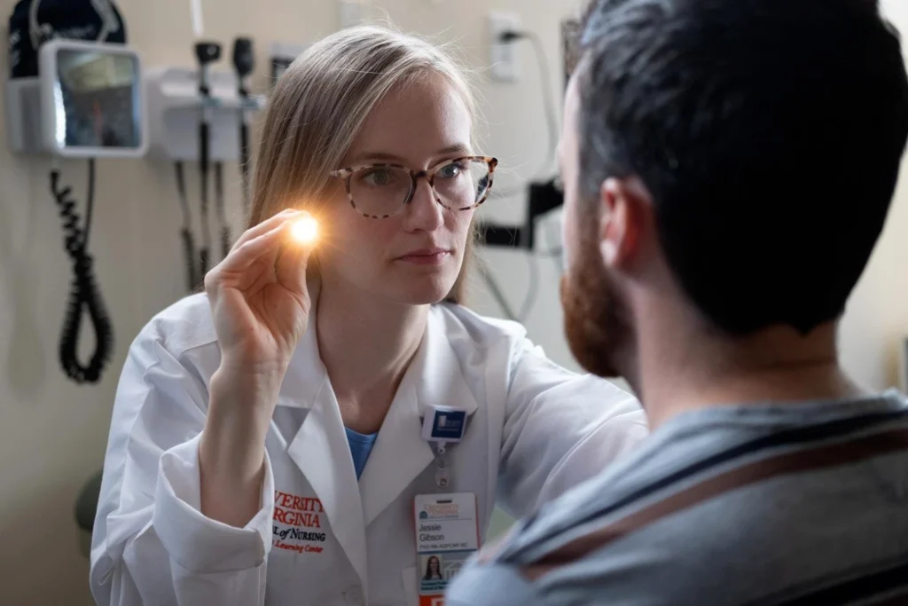
A neurological examination is an important component of clinical assessment and nursing practice. It involves observing the patient’s level of consciousness, examining the pupils, assessing cranial nerves, testing muscle strength and sensation, checking reflexes, and evaluating coordination and balance.
For nursing students, understanding the neurological examination is essential for recognising changes in a patient’s condition, documenting findings accurately and communicating concerns promptly to the healthcare team.
What Is a Neurological Examination?
A neurological examination is a structured clinical assessment of the brain, spinal cord, peripheral nerves and the functions they control. It helps healthcare professionals identify abnormalities that may be associated with neurological conditions, injuries or other medical problems.
The examination may be brief and focused or comprehensive, depending on the patient’s symptoms and clinical condition. For example, a patient with a sudden change in speech may require an urgent assessment for stroke, while someone attending a routine appointment for persistent numbness may undergo a more detailed examination of sensation and peripheral nerve function.
A neurological examination does not always establish a diagnosis on its own. The findings are interpreted alongside the patient’s medical history, vital signs, laboratory tests and imaging when required.
Why Is a Neurological Examination Important?
A neurological examination helps establish a baseline of nervous system function and identify changes that may require treatment or further investigation.
Its main purposes include:
- Assessing consciousness, alertness and orientation.
- Identifying weakness, paralysis or changes in muscle tone.
- Evaluating sensation, reflexes and coordination.
- Detecting possible signs of stroke or other neurological emergencies.
- Monitoring patients with head injuries or neurological disorders.
- Assessing changes in speech, vision, balance or memory.
- Evaluating progress during treatment and rehabilitation.
- Supporting clinical decision-making and care planning.
Repeated neurological assessments can be particularly important in patients whose condition may change rapidly. A new abnormal finding should be compared with previous observations and escalated according to the clinical setting and local protocol.
When Is a Neurological Examination Required?
A neurological examination may be indicated when a patient experiences:
- Sudden or persistent headaches.
- Weakness, numbness or tingling.
- Dizziness, loss of balance or coordination difficulties.
- Changes in vision, speech or swallowing.
- Confusion, reduced alertness or memory problems.
- Seizures or episodes of unexplained loss of consciousness.
- Suspected stroke or transient ischaemic attack.
- Head or spinal injuries.
- Symptoms associated with conditions such as Parkinson’s disease, multiple sclerosis or peripheral neuropathy.
The extent of the examination depends on the symptoms and the urgency of the situation. Sudden neurological changes require immediate assessment rather than waiting for a routine examination.
Types of Neurological Examination
The components selected for an assessment depend on the patient’s presentation.
1. Focused neurological examination
A focused examination evaluates the neurological functions most relevant to the patient’s symptoms. For example, a clinician may concentrate on facial movement, arm strength, speech and vision when assessing possible stroke.
2. Comprehensive neurological examination
A comprehensive examination assesses multiple areas, including mental status, cranial nerves, motor function, sensation, reflexes and coordination. It may also include gait, balance and specialised tests.
3. Serial neurological assessment
Serial assessment involves repeating selected observations at planned intervals. It is commonly used when monitoring patients with head injuries, altered consciousness or neurological conditions that may deteriorate.
4. Standardised consciousness assessment
Tools such as the Glasgow Coma Scale help clinicians describe a patient’s eye, verbal and motor responses. The tool is useful for structured communication but does not replace a complete neurological examination.
Preparation for a Neurological Examination
Good preparation supports accurate findings and helps the patient feel comfortable.
Before beginning, the healthcare professional should:
- Explain the purpose and main steps of the examination.
- Obtain consent and provide privacy.
- Review the presenting symptoms and relevant medical history.
- Ask about previous neurological conditions, medications and sensory difficulties.
- Gather necessary equipment, such as a penlight, reflex hammer, cotton wool and a suitable blunt or sharp sensory-testing instrument when indicated.
- Ensure adequate lighting and a safe environment.
- Assess whether pain, fatigue, language barriers or reduced consciousness may affect participation.
The examiner should use appropriate infection-prevention measures and adapt the examination to the patient’s abilities and clinical condition.
Step-by-Step Procedure of a Neurological Examination
A neurological examination follows a systematic approach. The following sections explain the major components commonly assessed in clinical practice.
1. Assessment of Mental Status and Consciousness
Begin by observing whether the patient is awake, alert and able to respond appropriately.
Assess orientation to person, place and time, as appropriate. Observe attention, memory, comprehension, speech and behaviour. Ask simple questions or give clear instructions suited to the patient’s language and abilities.
When consciousness is impaired, a standardised tool such as the Glasgow Coma Scale may be used according to clinical protocol. Document the findings and compare them with the patient’s baseline.
A new or worsening reduction in consciousness requires urgent assessment.
2. Examination of the Cranial Nerves
There are 12 pairs of cranial nerves, which contribute to functions such as smell, vision, eye movement, facial sensation, facial expression, hearing, swallowing and tongue movement.
A comprehensive cranial nerve examination may include:
- Olfactory nerve (I): Smell, when clinically indicated.
- Optic nerve (II): Visual acuity and visual fields, with additional assessment as appropriate.
- Oculomotor, trochlear and abducens nerves (III, IV and VI): Eye movements, eyelid position and pupil responses.
- Trigeminal nerve (V): Facial sensation and jaw movement.
- Facial nerve (VII): Facial expressions and symmetry.
- Vestibulocochlear nerve (VIII): Hearing and selected balance functions.
- Glossopharyngeal and vagus nerves (IX and X): Palatal movement, voice and swallowing-related functions, when appropriate.
- Accessory nerve (XI): Shoulder shrug and head rotation.
- Hypoglossal nerve (XII): Tongue movement.
The exact tests depend on the clinical situation. Suspected swallowing impairment requires appropriate safety assessment before oral intake is attempted.
3. Assessment of Motor Function
Motor assessment evaluates muscle strength, tone, movement and symmetry.
Ask the patient to perform selected movements against resistance, such as raising the arms, bending the elbows, extending the wrists or moving the legs. Compare corresponding muscle groups on both sides.
Muscle strength may be recorded using a standard grading scale, commonly from 0 to 5, where 0 indicates no visible or palpable contraction and 5 indicates normal power against resistance.
Observe for involuntary movements, muscle wasting, abnormal tone or asymmetry. New weakness, especially when sudden or one-sided, needs urgent assessment.
4. Assessment of Sensory Function
Sensory testing evaluates how the patient perceives different stimuli. Depending on the symptoms, this may include light touch, pain, temperature, vibration and joint position.
Explain each test and ask the patient to report what they feel. Compare corresponding areas on both sides of the body and assess whether the findings follow a consistent pattern.
Sensory testing should be performed carefully, using appropriate equipment and techniques. Avoid exposing the patient to harmful temperatures or using sharp instruments in a way that could cause injury.
5. Examination of Deep Tendon Reflexes
Reflex testing assesses responses that involve sensory nerves, the spinal cord and motor nerves.
A reflex hammer may be used to test selected deep tendon reflexes, such as the biceps, triceps, brachioradialis, patellar and Achilles reflexes.
Responses are commonly graded from absent to exaggerated, with the exact grading system specified in the clinical documentation. Reflexes should be compared between sides and interpreted alongside other findings.
Abnormal reflexes may suggest a neurological problem, but no single reflex result should be interpreted in isolation.
6. Assessment of Coordination
Coordination testing evaluates the ability to perform smooth, controlled movements. Common tests include finger-to-nose movement, rapid alternating hand movements and heel-to-shin testing.
Observe for tremor, inaccurate movement, slowness or difficulty coordinating the limbs. Findings may help clinicians assess cerebellar function and other motor pathways.
The tests should be adapted if the patient has pain, weakness or restricted movement.
7. Assessment of Gait and Balance
When it is safe to do so, observe the patient’s posture, walking pattern, step length, balance and ability to turn.
Additional tests may be used to assess balance, but they should only be performed when appropriate and with support to reduce the risk of falls. Patients who are dizzy, weak, confused or unstable should not be asked to walk without suitable assistance.
Document any difficulty walking, unusual gait patterns or need for support.
8. Assessment of Autonomic and Other Neurological Functions
When indicated, the examination may include assessment of autonomic functions, such as blood pressure changes with position, sweating abnormalities, bladder symptoms and bowel dysfunction.
The assessment may also consider speech, swallowing, pain patterns and other symptoms relevant to the suspected condition.
These findings should be interpreted in context, with additional investigations arranged when clinically necessary.
Equipment Used During a Neurological Examination
The equipment required depends on the examination being performed. Common items include:
- Penlight for pupil assessment.
- Reflex hammer for deep tendon reflexes.
- Visual acuity chart when vision testing is required.
- Cotton wool or other suitable tools for sensory assessment.
- A safe blunt or sharp sensory-testing instrument, when indicated.
- A tuning fork for selected vibration or hearing tests.
- A measuring device or appropriate monitoring equipment when clinically necessary.
- A documentation chart or electronic health record.
Not every examination requires all these items. The clinician should select equipment appropriate to the patient’s symptoms and the intended tests.
Benefits of a Neurological Examination
A neurological examination provides important clinical information without necessarily requiring specialised equipment or invasive procedures.
Its benefits include early identification of abnormal neurological signs, establishment of a baseline for comparison, support for diagnostic decisions and monitoring of changes during treatment.
It can also help guide decisions about further investigations, specialist referral, rehabilitation and patient safety measures.
However, a normal examination does not rule out every neurological condition. Some disorders require imaging, blood tests, electrophysiological studies or other investigations.
Risks and Limitations
A routine neurological examination is generally non-invasive and low risk. Nevertheless, patient safety and clinical judgement remain essential.
Some balance tests can increase the risk of falls, and sensory tests must be performed safely. Pain, anxiety, medication effects, fatigue, language barriers and impaired consciousness may influence the results.
The examination may also be limited by the patient’s ability to cooperate or by the time available during an emergency. Findings must therefore be interpreted alongside the full clinical picture.
Nursing Responsibilities During a Neurological Examination
Nurses contribute to neurological assessment through observation, measurement, documentation, patient education and timely escalation.
Key responsibilities include:
- Establishing the patient’s baseline neurological status.
- Monitoring consciousness and other observations at the prescribed frequency.
- Assessing pupils and limb movement when required by the care plan and the nurse’s training.
- Recognising new weakness, altered speech, unequal pupils or changes in consciousness.
- Maintaining patient safety during movement and balance assessment.
- Recording findings accurately, including the time of assessment.
- Comparing new findings with previous observations.
- Reporting deterioration promptly to the responsible clinician.
- Supporting the patient and explaining procedures clearly.
- Following local protocols for urgent neurological changes.
Nurses should document objective findings rather than relying on vague descriptions. For example, describing which limb is weak and what movement the patient can perform is more useful than simply recording that the patient appears unwell.
What Should Be Documented?
Accurate documentation supports continuity of care and helps clinicians identify changes over time.
Depending on the assessment, records may include:
- Level of consciousness and relevant consciousness score.
- Orientation, speech and ability to follow commands.
- Pupil size, symmetry and reaction to light.
- Muscle strength and symmetry of movement.
- Sensory findings and reflex responses.
- Coordination, gait and balance, if assessed.
- Symptoms reported by the patient.
- Time of assessment and any changes from baseline.
- Actions taken, the clinician notified and the patient’s response.
Documentation should follow the clinical facility’s approved charting system and escalation policy.
When Is a Neurological Examination an Emergency?
Some neurological symptoms require immediate emergency evaluation.
Call for emergency medical assistance if a person develops sudden facial drooping, weakness or numbness on one side, difficulty speaking, sudden loss of vision, severe sudden headache, a seizure lasting longer than expected, or a new reduction in consciousness.
These symptoms can indicate a stroke or another serious condition. Do not delay emergency care while attempting to complete a routine neurological examination.
For possible stroke, note the time symptoms began or the last time the person was known to be well, if that information is available.
Why Is Neurological Examination Important for Nursing Students?
Neurological examination is a core clinical skill for nursing students because it develops observation, assessment, documentation and clinical reasoning.
Students need to understand the purpose of each component, recognise normal and abnormal findings, and know when to seek assistance. Practical skills should be developed under qualified supervision and according to the student’s scope of practice.
At Ideal Institute of Nursing, learning neurological assessment can help students connect anatomy and physiology with medical-surgical nursing, emergency care and rehabilitation. Simulation, supervised clinical practice and structured feedback can strengthen their confidence and accuracy.
Frequently Asked Questions About Neurological Examination
1. What is a neurological examination?
A neurological examination is a structured assessment of nervous system function, including consciousness, cranial nerves, muscle strength, sensation, reflexes, coordination and balance.
2. Why is a neurological examination performed?
It helps identify abnormalities that may be associated with stroke, head injury, nerve disorders, seizures or other neurological conditions. It can also establish a baseline and monitor changes over time.
3. What are the main components of a neurological examination?
The main components are mental status and consciousness, cranial nerve function, motor assessment, sensory testing, reflexes, coordination and gait or balance when appropriate.
4. What equipment is used in a neurological examination?
Common equipment includes a penlight, reflex hammer, visual acuity chart, sensory-testing materials and a tuning fork for selected tests. The equipment depends on the patient’s symptoms and the scope of the examination.
5. How is muscle strength assessed?
The examiner asks the patient to perform selected movements against resistance and compares both sides. Strength is often graded on a 0-to-5 scale, according to the assessment system used.
6. What does the Glasgow Coma Scale assess?
The Glasgow Coma Scale records eye, verbal and motor responses to help describe a person’s level of consciousness. It is one component of neurological assessment, not a substitute for a full examination.
7. How often should neurological observations be performed?
The frequency depends on the patient’s condition, clinical orders and local protocol. Patients with acute neurological conditions or head injuries may need frequent repeated observations.
8. What abnormal findings require urgent attention?
Sudden weakness, facial drooping, speech difficulties, a new reduction in consciousness, unequal pupils with acute symptoms, or a sudden severe headache may indicate a medical emergency. Immediate assessment and escalation are essential.
9. What is the nurse’s role in neurological examination?
Nurses monitor and document neurological status, recognise changes from baseline, maintain safety, communicate concerns promptly and carry out assessments within their training and scope of practice.
Conclusion
Neurological examination is a fundamental clinical assessment that helps healthcare professionals evaluate the functioning of the brain, spinal cord and peripheral nerves. By systematically assessing consciousness, cranial nerves, motor strength, sensation, reflexes, coordination and balance, clinicians can identify important abnormalities and monitor changes over time.
For nursing students, learning the neurological examination develops essential clinical observation, documentation and communication skills. Accurate assessment, patient safety and prompt escalation of concerning findings are central to effective neurological care.
Educational disclaimer: This article is intended for nursing education and general information. It does not replace clinical training, professional judgement, local protocols or emergency medical assessment.